SCS Science Image Challenge 2012 Winner
Department of Physics and Beckman Institute. Theoretical and computational biophysics group. Glasswork by Luke Jerram.
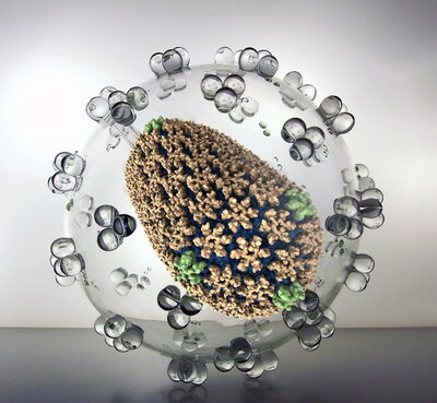
Mature HIV virus capsid
Viral particles of HIV-1 contain conical cores (or "cones") formed by a protein shell composed of the viral capsid protein (CA). The capsid protein (CA) plays critical roles in both late and early stages of the infection process and is widely viewed as an important unexploited therapeutic target. Capsid protein assembles into hexamers (gold) and pentamers (green), the curvature of the capsid is determined by the location of the pentamers.
Finalist
Department of Chemistry
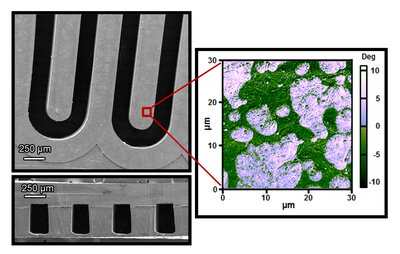
Disposable micro gas chromatograph
A phase-separated polymer microchannel acts as an inexpensive and disposable micro gas chromatograph with no separately-coated stationary phase. SEMs of unsealed (top left) and sealed (bottom left, cross-section) polymer microchannel fabricated via a replica molding process are shown; phase contrast AFM (right) shows two domains at the polymer surface.
Finalist
Department of Chemistry
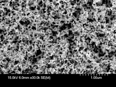
Etched nanoporous gold
Scanning electron micrograph of a 250 µm thick 10 karat white gold sheet after immersion in concentrated nitric acid for 2 days followed by dipping in aqua regia for 1 minute to remove less noble metals and create a porous structure. Aqua regia dissolves and thins the gold ligaments.
Finalist
TCBG, Department of Physics
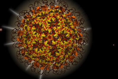
Nudaurelia capensis ω virus
The maturation process of Nudaurelia capensis ω Virus (NωV) is depicted by overlaying several cryo-EM density maps and an all-atom model of the virus capsid at different maturation stages.
Finalist
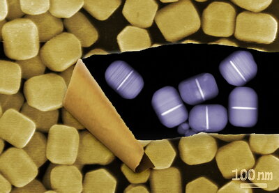
Department of Chemistry
Hexagonal rods formed from DNA-directed colloidal reduction of silver on gold plates
Hexagonal rods formed from DNA-directed colloidal reduction of silver on gold plates. The outer image, obtained via scanning electron microscopy, shows the overall morphology of the particle. This image is torn back, uncovering a scanning transmission electron micrograph of the same particles, revealing the gold plates (bright regions) hidden inside.