
Department of Chemistry
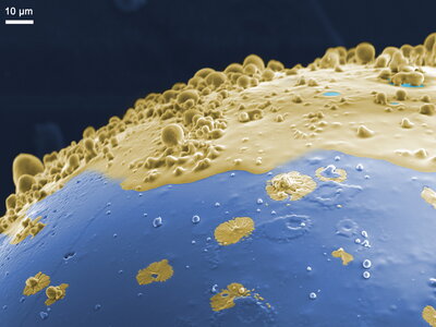
From the New World: A Sugar Planet
This is a false-colored SEM image showing the morphological features developed on the surface of a sucrose crystal that is caramelized under ultrasonic irradiation.
Department of Chemistry
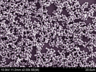
Peanut hematite particles
Scanning electron micrograph of weakly ferromagnetic hematite crystal in peanut shape. The highly asymmetric morphology is obtained by aging the particle in sodium sulfate to modulate its facets.
Department of TCBG, Department of Physics and Beckman Institute
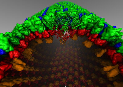
The atomic model of the immature retroviral lattice of Rous Sarcoma Virus shown here consists of 300 hexamers arranged in a tubular lattice. The central hexamer is highlighted at higher structural details. The figure was produced solely by using VMD (Visual Molecular Dynamics).
Department of ChBE
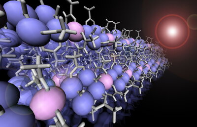
Infinite PdPt Bimetallic Chain
Infinite PdPt bimetallic chain crystal was synthesized. This infinite chain structure is based on a new motif with alternatively connected Pd4(CO)4(OAc)4 paddlewheel molecules and Pt(acac)2 square-planar molecules. In this crystal structure, Pd (blue) and Pt (pink)atoms are closely packed in to one dimensional metal atom wires.
Department of Physics
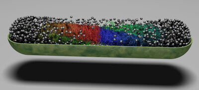
Cross-section of E. coli cell showing the conformation of the genome from Brownian Dynamics simulations. Ribosomes are represented by gray spheres. This geometry is used as input to our Lattice Microbe simulations of whole-cell processes. Image produced using PovRay/Python3 workflow.