2015 SCS Science Image Challenge Winner
Department of Chemistry
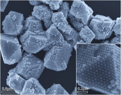
Palladium Supercrystals
SEM image of supercrystals formed by palladium nano-cuboctahedron monomers. These crystals are formed by the self-assembly of the monomers due to the slow diffusion of surfactant (cetrimonium chloride) into a concentrated colloidal solution of palladium nano-cuboctahedron.The supercrystals formed are highly dependent on the shape of the monomer nanoparticle used.
Finalist
Department of Chemical and Biomolecular Engineering
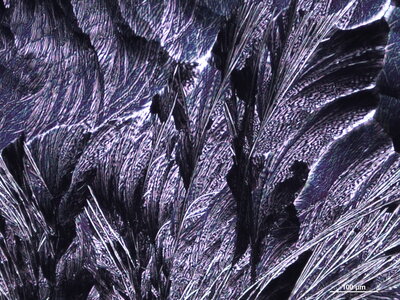
Beauty in the Curve
Curved aspirin crystallites are observed in this cross polarized optical micrograph. The curvature in the crystallites is observed when a two-step mechanism of solution deposition and subsequent spherulite growth occurs during solution printing on an SiO2 wafer.
Finalist
Department of Chemistry
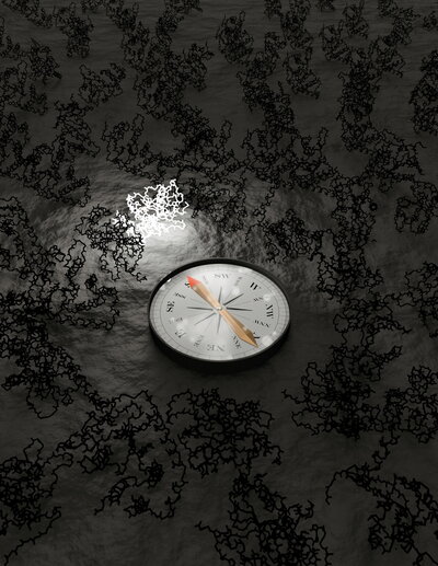
COMPASS: Navigating the Protein Landscape
NMR spectroscopy is a powerful technique for determining protein structures that traditionally requires months of data analysis. COMPASS replaces manual data analysis with a computer vision-inspired algorithm that identifies the unique structure using a single spectrum. This artistic representation depicts COMPASS revealing the correct fold in the dark structural landscape.
Finalist
Department of Chemistry
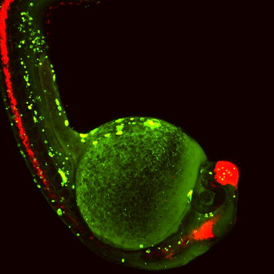
Rapid Apoptosis in Zebrafish
Apoptotic cell death in transgenic YFP zebrafish embryos using an anti-cancer small molecule.
Finalist
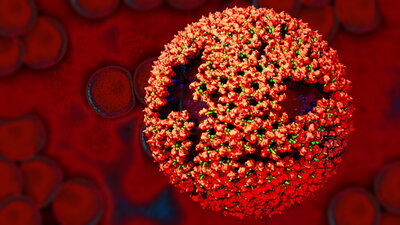
Imperfect intruder
An immature retrovirus has a near-spherical capsid with plenty of structural defects and is therefore non-infectious. The blood cells at the background illustrate the viruses (blue dots) successful entered the cells but failed to hijack the cell’s machinery. The figure of the retrovirus was produced using VMD (Visual Molecular Dynamics).